top of page


Cone Beam CT Scanner
Advanced 3D Imaging for Precise Endodontic Care
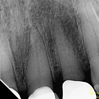
Our practice uses advanced Cone-Beam CT (computed tomography) technology to capture highly detailed 3D images for precise diagnosis, planning, and treatment of endodontic conditions. This state-of-the-art imaging allows us to visualize teeth, bone, sinuses, and surrounding structures with exceptional accuracy—while minimizing radiation exposure. By providing a clearer, more comprehensive view than traditional 2D dental X-rays, we ensure the highest level of precision and patient care. Often we are able to identify problems that would be routinely missed on the traditional periapical image as seen to the left.


bottom of page